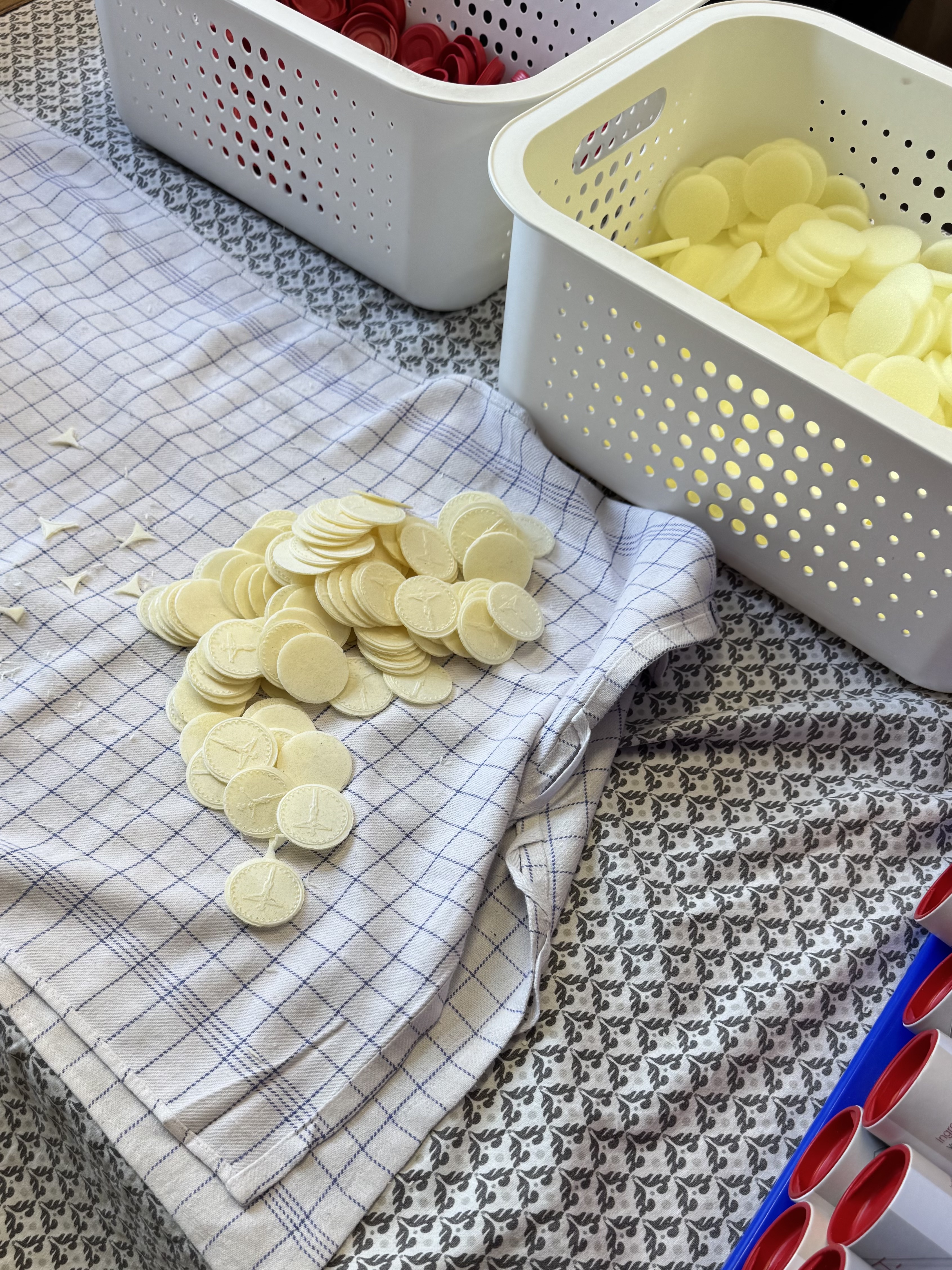

When a wafer consecrated at Mass develops a red patch, a photograph can seem to tell the whole story. To Catholics, the object is sacred; a stain resembling blood may look like a visible confirmation of Christ’s presence. But a photograph cannot identify the stain. That takes a sample, a record of its handling and tests that distinguish blood from materials that resemble it.
A study published on 5 March 2025 offers a useful starting point. Tomasz Grzybowski and colleagues examined 25 anonymized cases referred by Church officials, using combinations of microscopy, microbial investigation, blood tests and DNA analysis. The work proposes an investigative procedure; it does not describe a uniform battery of tests applied to every case. [1]
The useful question is how the researchers worked from appearance to identification. “Red,” “blood,” “human” and “miraculous” each require their own evidence.
A positive screen starts a question
Forensic laboratories distinguish screening from identification. A screening test looks for an indication that a substance may be present. It can be useful precisely because it detects small amounts, yet its reaction may also be produced by other materials. A positive result can justify further work without supplying the final identification. [2] [3]
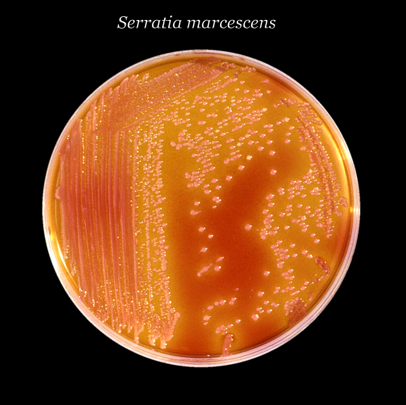
In the 2025 study, one screen relied on the chemical activity of haemoglobin, the oxygen-carrying protein in blood. Microbial enzymes can trigger the same indicator reaction. The authors identify this as a plausible source of false positives in contaminated specimens. The test detects a reaction; further tests must identify what produced it. [1]
The National Institute of Justice illustrates the distinction with a hypothetical stain that tests presumptively positive for blood and yields a DNA profile matching a person. Those results alone do not establish that the stain is that person’s blood. The DNA result answers a genetic question; the missing confirmation concerns what bodily material is present. Combining two impressive findings cannot replace the missing step. [2]
Negative results need equally careful language. The U.S. Department of Justice’s guidance for its forensic examiners distinguishes material not detected from a claim that no material could possibly be there. Too little biological material, or material in poor condition, can limit detection. The reader should therefore ask what the test detects and under what conditions it works. [3]

DNA needs a history
Modern DNA methods can detect material from very few skin cells. That sensitivity is powerful, but it also makes an object’s handling history important. NIST, the U.S. National Institute of Standards and Technology, explains that DNA can move between surfaces, including through indirect transfer. Finding a trace does not by itself establish when or how it arrived. [4]
Consider the difference between identifying a possible source and reconstructing an event. A genetic result may support the first while leaving the second uncertain. NIST’s scientific review of DNA mixtures emphasizes that interpreting the activity that deposited DNA requires context beyond the profile itself. The same distinction matters whenever a tiny trace carries a large historical or religious claim. [5]
For a host investigation, that suggests practical questions for the record: who handled the material, what container held it, when was it sampled, and what happened before it reached the laboratory? These are questions to establish provenance. That record helps distinguish a trace present before the event from one introduced during collection or examination. [5]
What the 2025 cases leave us with
The researchers identified bacteria and fungi in several specimens. They also reported isolated skin cells and red blood cells in case 8, and a small amount of human DNA in case 21, which they interpreted as possibly transferred through handling. A blanket statement that no human material appeared would lose these qualifications. [1]
The difficult specimens deserve a closer look. In case 1, microscopy showed reddish-brown bundles with striations and nuclei resembling cardiac muscle; a fungal stain was negative. Case 2 looked similar, but lacked visible nuclei, and antibody-based testing did not identify muscle tissue. The authors left both for further investigation. These conflicting observations do not confirm cardiac tissue. The anonymized cases also cannot be assigned to familiar shrines or used to settle every reported Eucharistic phenomenon. [1]
The unresolved specimens make a demanding test of any account of this research. A summary should include them alongside the microbial findings, then ask what analysis of the material could resolve the conflicting results. A striking resemblance is a reason to investigate further.
A local investigation, a limited public record
A separate example shows what an institutional conclusion can look like. In March 2025, the Archdiocese of Indianapolis reported testing a discolored host from St. Anthony Church in Morris, Indiana. Its newspaper, The Criterion, relayed a 24 March statement describing fungus and three bacterial species commonly found on hands; it reported that no human blood was detected. The discoloration had been observed in February. [6]
The newspaper provides the archdiocese’s conclusion without the complete laboratory report. We can report that conclusion, but cannot check the underlying measurements from this public account. The Indiana case is separate from the anonymized research series. [6]
What Catholic belief is claiming
Catholic teaching gives this inquiry a particular context. Paragraph 1376 of the Catechism describes transubstantiation as the conversion of the whole substance of bread and wine into Christ’s body and blood. Paragraph 1381 presents Christ’s presence as apprehended by faith rather than by the senses. The Church’s Compendium adds that the outward characteristics of bread and wine remain. This teaching does not predict a visible biological transformation at every Mass. [7] [8]
A reported change of color or texture adds a physical claim to that sacramental belief. A laboratory can investigate the stain’s composition and possible causes. For a Catholic, discovering a microbial cause would explain that visible change without settling the doctrine about Christ’s sacramental presence. Someone who does not share the doctrine can still assess the same laboratory evidence. [7]
Before sharing a photograph of a changed host, look for the observation behind the claim: what was sampled, which tests were performed, and what the results showed. Respect for the sacred object includes care for the accuracy of its story.